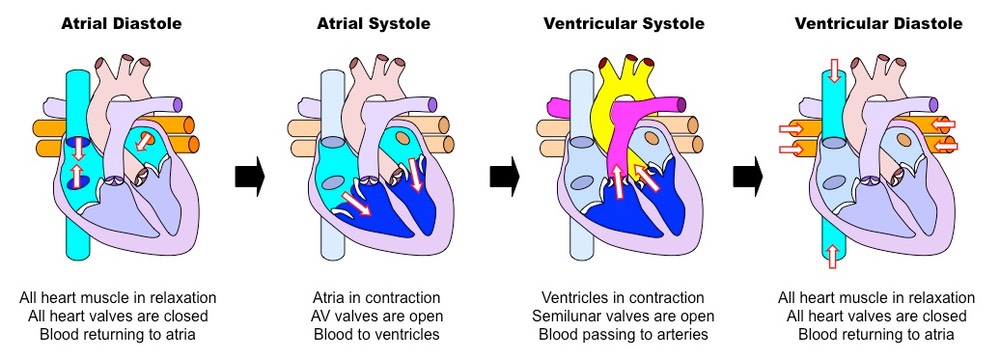
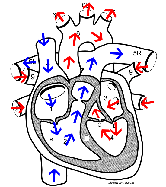
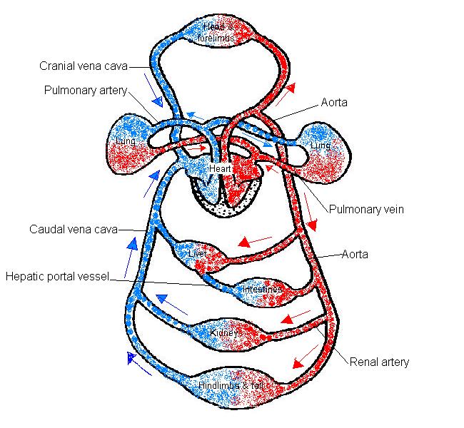
43 heart diagram with labels and blood flow
A& P II Chapter 16 Lab Flashcards | Quizlet Drag each label to the appropriate location on the diagram of a homeostasis pathway. Check the picture for answer. Which structure is highlighted? ... increased heart rate increased blood sugar level decreased blood sugar level ... Hypothalamic hormones flow in blood through portal veins 4) Stimulation or inhibition of hormone secretion by ... Heart Information Center: Heart Anatomy - Texas Heart Institute The left ventricle's chamber walls are only about a half-inch thick, but they have enough force to push blood through the aortic valve and into your body. The Heart Valves. Four valves regulate blood flow through your heart: The tricuspid valve regulates blood flow between the right atrium and right ventricle.
(PDF) Heart Disease Prediction System - ResearchGate different medical attributes such as blood sugar and heart rat e, age, ... Data Flow Diagram. Datas et Pre processing. ... handling numerous class labels in the prediction process, and it …
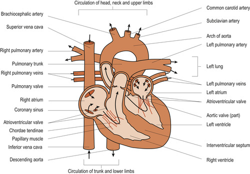
Heart diagram with labels and blood flow
Diagram of Blood Flow Through the Heart - Bodytomy The heart is divided into two chambers, left and right, the right atrium and ventricle lie on the right side and the left atrium and ventricle on the left side. These two chambers are not directly connected to each other. Synchronization of the Two Chamber The right and left side or chambers of the heart work in tandem with each other. Human Heart - Diagram and Anatomy of the Heart - Innerbody The heart is a muscular organ about the size of a closed fist that functions as the body's circulatory pump. It takes in deoxygenated blood through the veins and delivers it to the lungs for oxygenation before pumping it into the various arteries (which provide oxygen and nutrients to body tissues by transporting the blood throughout the body). YR 8 Topic 2 Circulatory System - Pinterest Label these heart parts. Jody Peck. Science. Human Anatomy Chart. ... Diagram of Coronary heart Blood Movement for Cardiac Nursing College students. ... Student Nurse. A&P 2 Chapter 19&20 The Circulatory System: Heart and Blood Vessels Flashcards | Quizlet. Nursecepts | Nursing Student Resource | Nursing School Resource. Nursing Cardiovascular ...
Heart diagram with labels and blood flow. Heart Diagram with Labels and Detailed Explanation - BYJUS Diagram of Heart. The human heart is the most crucial organ of the human body. It pumps blood from the heart to different parts of the body and back to the heart. The most common heart attack symptoms or warning signs are chest pain, breathlessness, nausea, sweating etc. The diagram of heart is beneficial for Class 10 and 12 and is frequently ... Blood Flow Diagram Pictures, Images and Stock Photos Browse 2,626 blood flow diagram stock photos and images available, or search for heart blood flow diagram to find more great stock photos and pictures. A simplified diagram showing blood circulation in the human body. A simple diagram of the human body with the blood flow, heart and lungs shown, graphically but accurately. › 1-label-the-heartLabel the heart — Science Learning Hub Label the heart Interactive Add to collection In this interactive, you can label parts of the human heart. Drag and drop the text labels onto the boxes next to the diagram. Selecting or hovering over a box will highlight each area in the diagram. Right ventricle Right atrium Left atrium Pulmonary artery Left ventricle Pulmonary vein Semilunar valve Labelling the heart — Science Learning Hub Blood transports oxygen and nutrients to the body. It is also involved in the removal of metabolic wastes. In this interactive, you can label parts of the human heart. Drag and drop the text labels onto the boxes next to the diagram. Selecting or hovering over a box will highlight each area in the diagram.
Human Heart Diagram Labeled | Science Trends Let's examine the anatomy of the heart along with some diagrams that show how the heart operates. Anatomy Of The Heart The human heart usually weighs somewhere between 10 to 12 ounces in men and between 8 to 10 ounces in women, and in terms of size is roughly the size of the fist. Label the heart — Science Learning Hub Jun 16, 2017 · Labels. Description. Vena cava. Carries deoxygenated blood from the body to the heart. Semilunar valve. Flaps that prevent backflow of blood. Left atrium. Receives oxygenated blood from the lungs. Left ventricle. Region of the heart that pumps oxygenated blood to the body. Pulmonary artery. Carries deoxygenated blood to the lungs. Right ventricle › photos › muscular-systemMuscular System Labeled Diagram Stock Photos, Pictures ... Heart blood flow circulation anatomical diagram with atrium and ventricle system. Vector illustration labeled medical poster. Heart blood flow anatomical diagram with atrium and ventricle system. Vector illustration labeled medical poster. Blood circulation path scheme with arrows. muscular system labeled diagram stock illustrations The Heart | Circulatory Anatomy - Visible Body One chamber on the left receives oxygen-rich blood from the lungs and another pumps that nutrient-rich blood into the body. Two valves control blood flow within the heart's chambers, and two valves control blood flow out of the heart. 1. The Heart Wall Is Composed of Three Layers. The muscular wall of the heart has three layers.
Label Heart Anatomy Diagram Printout - EnchantedLearning.com The blood is then pumped to the left ventricle, then the blood is pumped through the aorta and to the rest of the body. This cycle is then repeated. Every day, the heart pumps about 2,000 gallons (7,600 liters) of blood, beating about 100,000 times. Label the heart anatomy diagram below using the heart glossary. Note: On the diagram, the right ... Circulatory System Diagram - New Health Advisor This circuit typically includes the movement of blood inside heart and 'myocardium' (the membrane of heart). Coronary circuit mainly consists of cardiac veins including anterior cardiac vein, small vein, middle vein and great (large) cardiac vein. There are different types of circulatory system diagrams; some have labels while others don't. pmt.physicsandmathstutor.com › download › BiologyPractical notes - SP 2.3c Dissection of a Mammalian Heart ... 3. Use a glass rod to follow the path of blood flow : via the pulmonary vein, left atrium and through the bicuspid valve into the left ventricle; via the left ventricle through the semilunar valve and out of the aorta. 4. Note the muscular surface of the ventricle chambers which ensur es smooth blood flow. 5. PDF BLOOD FLOW THROUGH THE HEART diagram deoxygenated blood from body tissue oxygenated blood from lungs via pulmonary vein s superior and inferior vena cava left atrium right atrium bicuspid valve tricuspid valve left ventricle ... blood flow through the heart diagram author: taustin created date: 9/30/2013 9:09:13 pm
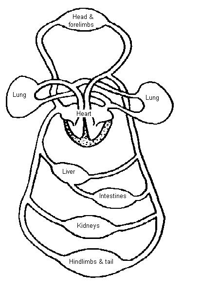
Circulatory System Diagram - Cardiovascular System and Blood ... The veins of the systemic circulation system take the then deoxygenated blood from the body back to the heart. Types of Circulatory System Diagrams. There are several different circulatory system diagrams. They may come with or without labels. Common circulatory system diagrams show pulmonary circulation, coronary circulation, systematic ...
byjus.com › biology › human-heartHuman Heart - Anatomy, Functions and Facts about Heart The external structure of the heart has many blood vessels that form a network, with other major vessels emerging from within the structure. The blood vessels typically comprise the following: Veins supply deoxygenated blood to the heart via inferior and superior vena cava, and it eventually drains into the right atrium.
Free Blank Heart Diagram, Download Free Blank Heart Diagram png images, Free ClipArts on Clipart ...
Practical notes - SP 2.3c Dissection of a Mammalian Heart 3. Use a glass rod to follow the path of blood flow : via the pulmonary vein, left atrium and through the bicuspid valve into the left ventricle; via the left ventricle through the semilunar valve and out of the aorta. 4. Note the muscular surface of the ventricle chambers which ensur es smooth blood flow. 5.
Anatomy of the Heart: Blood Flow and Parts - Study.com The Heart. In your body, blood flows within a closed circuit of blood vessels. Blood is able to circulate around your body thanks to a muscular pump known as your heart. As we previously learned ...

Easy-to-follow arrows for blood flow #Cardiac #MedSurg #Nursing | Nursing | Médical, Anatomie ...
› pmc › articlesCerebral blood flow and autoregulation: current measurement ... Jun 21, 2016 · 1.1. Physiological importance and normal values of cerebral blood flow in adult humans. The human brain is an organ with high-energy density demands, amounting to only 2% of the entire body mass (or ∼ 1.4 kg) but accounting for about 20% of the total power consumption of a normal adult at rest (or ∼ 20 W).
Heart anatomy: Structure, valves, coronary vessels - Kenhub The cusps are pushed open to allow blood flow in one direction, and then closed to seal the orifices and prevent the backflow of blood. Backward prolapse of the cusps is prevented by the chordae tendineae-also known as the heart strings-fibrous cords that connect the papillary muscles of the ventricular wall to the atrioventricular valves.. There are two sets of valves: atrioventricular ...
File:Heart diagram blood flow en.svg - Wikipedia File:Heart diagram blood flow en.svg. Size of this PNG preview of this SVG file: 330 × 370 pixels. Other resolutions: 214 × 240 pixels | 428 × 480 pixels | 535 × 600 pixels | 685 × 768 pixels | 913 × 1,024 pixels | 1,827 × 2,048 pixels. This is a file from the Wikimedia Commons. Information from its description page there is shown below.
The Heart Year 6 | KS2 Science | Twinkl (teacher made) Follow your heart and download this Animals Including Humans science lesson pack. This engaging resource enables children to learn about the three parts of the circulatory system and most specifically, the role of the heart. In this lesson, children recap on the names of organs that they have learnt in previous years and then they go on to learn about how the heart, blood …
A Diagram of the Heart and Its Functioning Explained in Detail The heart blood flow diagram (flowchart) given below will help you to understand the pathway of blood through the heart.Initial five points denotes impure or deoxygenated blood and the last five points denotes pure or oxygenated blood. 1.Different Parts of the Body. ↓. 2.Major Veins.
Muscular System Labeled Diagram Pictures, Images and Stock … Frontal view of the muscular system of the male human body with descriptive labels pointing to the muscles on a white background. ... Heart blood flow anatomical diagram with atrium and ventricle system. Vector illustration labeled medical poster. Blood circulation path scheme with arrows. muscular system labeled diagram stock illustrations.
ANS-HEART LABEL AND BLOOD FLOW .docx - Circulation Exercise... View ANS-HEART LABEL AND BLOOD FLOW .docx from MGMT MISC at Wayne County Community College District. Circulation Exercise STRUCTURE OF THE HUMAN HEART: using your textbook or notes, label the ... label the following diagram of the human heart. A. superior vena cava L B. right atrium C. tricuspid valve A K D right ventricle . E. pulmonary valve ...
Dopamine - Wikipedia Dopamine (DA, a contraction of 3,4-dihydroxyphenethylamine) is a neuromodulatory molecule that plays several important roles in cells.It is an organic chemical of the catecholamine and phenethylamine families. Dopamine constitutes about 80% of the catecholamine content in the brain. It is an amine synthesized by removing a carboxyl group from a molecule of its precursor …
PDF Heart Diagram Answer Key - University of Washington A: om the body: t lung VEINS: trium VENTRICLE: o the lung TRIUM VE A: o the body: o the lungs APEX VENTRICLE: o the body all VE TRIUM VEINS: trium e e A: om the body
Heart Blood Flow | Simple Anatomy Diagram, Cardiac Circulation Pathway ... Step 1 and 6 involve a blood vessel, which makes sense as this is how blood enters and exits that side of the heart. Steps 2-5 involve a chamber, valve, chamber, and valve. So if you remember this general pattern, it will help you recall the order in which blood flows through each side of the heart.
Human Heart - Anatomy, Functions and Facts about Heart Myocardial infarction is a serious medical condition where the blood flow to the heart is reduced or entirely stopped. This causes oxygen deprivation in the heart muscles, and prolonged deprivation can cause tissues to die. ... Label the Heart Diagram below: Practice your understanding of the heart structure. Drag and drop the correct labels to ...
Cerebral blood flow and autoregulation: current measurement techniques ... Jun 21, 2016 · 1.1. Physiological importance and normal values of cerebral blood flow in adult humans. The human brain is an organ with high-energy density demands, amounting to only 2% of the entire body mass (or ∼ 1.4 kg) but accounting for about 20% of the total power consumption of a normal adult at rest (or ∼ 20 W).Blood perfusion is responsible for the delivery of oxygen, …
en.wikipedia.org › wiki › DopamineDopamine - Wikipedia Its effects, depending on dosage, include an increase in sodium excretion by the kidneys, an increase in urine output, an increase in heart rate, and an increase in blood pressure. At low doses it acts through the sympathetic nervous system to increase heart muscle contraction force and heart rate, thereby increasing cardiac output and blood ...





Post a Comment for "43 heart diagram with labels and blood flow"